Target Information
| Target General Information | Top | |||||
|---|---|---|---|---|---|---|
| Target ID |
T47623
(Former ID: TTDC00060)
|
|||||
| Target Name |
Heparanase (HPSE)
|
|||||
| Synonyms |
Hpa1; Heparanase-1; Heparanase 8 kDa subunit; Heparanase 50 kDa subunit; HSE1; HPSE1; HPR1; HPA; HEP protein; Endo-glucoronidase
Click to Show/Hide
|
|||||
| Gene Name |
HPSE
|
|||||
| Target Type |
Successful target
|
[1] | ||||
| Disease | [+] 2 Target-related Diseases | + | ||||
| 1 | African trypanosomiasis [ICD-11: 1F51] | |||||
| 2 | Ocular disease [ICD-11: N.A.] | |||||
| Function |
Participates in extracellular matrix (ECM) degradation and remodeling. Selectively cleaves the linkage between a glucuronic acid unit and an N-sulfo glucosamine unit carrying either a 3-O-sulfo or a 6-O-sulfo group. Can also cleave the linkage between a glucuronic acid unit and an N-sulfo glucosamine unit carrying a 2-O-sulfo group, but not linkages between a glucuronic acid unit and a 2-O-sulfated iduronic acid moiety. It is essentially inactive at neutral pH but becomes active under acidic conditions such as during tumor invasion and in inflammatory processes. Facilitates cell migration associated with metastasis, wound healing and inflammation. Enhances shedding of syndecans, and increases endothelial invasion and angiogenesis in myelomas. Acts as procoagulant by increasing the generation of activation factor X in the presence of tissue factor and activation factor VII. Increases cell adhesion to the extracellular matrix (ECM), independent of its enzymatic activity. Induces AKT1/PKB phosphorylation via lipid rafts increasing cell mobility and invasion. Heparin increases this AKT1/PKB activation. Regulates osteogenesis. Enhances angiogenesis through up-regulation of SRC-mediated activation of VEGF. Implicated in hair follicle inner root sheath differentiation and hair homeostasis. Endoglycosidase that cleaves heparan sulfate proteoglycans (HSPGs) into heparan sulfate side chains and core proteoglycans.
Click to Show/Hide
|
|||||
| BioChemical Class |
Glycosylase
|
|||||
| UniProt ID | ||||||
| EC Number |
EC 3.2.1.166
|
|||||
| Sequence |
MLLRSKPALPPPLMLLLLGPLGPLSPGALPRPAQAQDVVDLDFFTQEPLHLVSPSFLSVT
IDANLATDPRFLILLGSPKLRTLARGLSPAYLRFGGTKTDFLIFDPKKESTFEERSYWQS QVNQDICKYGSIPPDVEEKLRLEWPYQEQLLLREHYQKKFKNSTYSRSSVDVLYTFANCS GLDLIFGLNALLRTADLQWNSSNAQLLLDYCSSKGYNISWELGNEPNSFLKKADIFINGS QLGEDFIQLHKLLRKSTFKNAKLYGPDVGQPRRKTAKMLKSFLKAGGEVIDSVTWHHYYL NGRTATKEDFLNPDVLDIFISSVQKVFQVVESTRPGKKVWLGETSSAYGGGAPLLSDTFA AGFMWLDKLGLSARMGIEVVMRQVFFGAGNYHLVDENFDPLPDYWLSLLFKKLVGTKVLM ASVQGSKRRKLRVYLHCTNTDNPRYKEGDLTLYAINLHNVTKYLRLPYPFSNKQVDKYLL RPLGPHGLLSKSVQLNGLTLKMVDDQTLPPLMEKPLRPGSSLGLPAFSYSFFVIRNAKVA ACI Click to Show/Hide
|
|||||
| 3D Structure | Click to Show 3D Structure of This Target | PDB | ||||
| HIT2.0 ID | T14Q85 | |||||
| Drugs and Modes of Action | Top | |||||
|---|---|---|---|---|---|---|
| Clinical Trial Drug(s) | [+] 2 Clinical Trial Drugs | + | ||||
| 1 | PG-545 | Drug Info | Phase 1 | Ocular disease | [2] | |
| 2 | Suramin | Drug Info | Phase 1 | African trypanosomiasis | [1], [3] | |
| Discontinued Drug(s) | [+] 1 Discontinued Drugs | + | ||||
| 1 | Neutralase | Drug Info | Discontinued in Phase 3 | Angiogenesis disorder | [4] | |
| Mode of Action | [+] 2 Modes of Action | + | ||||
| Modulator | [+] 2 Modulator drugs | + | ||||
| 1 | PG-545 | Drug Info | [2] | |||
| 2 | Neutralase | Drug Info | [5] | |||
| Inhibitor | [+] 1 Inhibitor drugs | + | ||||
| 1 | Suramin | Drug Info | [1], [3] | |||
| Cell-based Target Expression Variations | Top | |||||
|---|---|---|---|---|---|---|
| Cell-based Target Expression Variations | ||||||
| Drug Binding Sites of Target | Top | |||||
|---|---|---|---|---|---|---|
| Ligand Name: 4-Nitrophenol | Ligand Info | |||||
| Structure Description | Glycoside Hydrolase ligand structure 1 | PDB:5E97 | ||||
| Method | X-ray diffraction | Resolution | 1.63 Å | Mutation | No | [6] |
| PDB Sequence |
> Chain A
KFKNSTYSRS 168 SVDVLYTFAN178 CSGLDLIFGL188 NALLRTADLQ198 WNSSNAQLLL208 DYCSSKGYNI 218 SWELGNEPNS228 FLKKADIFIN238 GSQLGEDFIQ248 LHKLLRKSTF258 KNAKLYGPDV 268 GQPRRKTAKM278 LKSFLKAGGE288 VIDSVTWHHY298 YLNGRTATRE308 DFLNPDVLDI 318 FISSVQKVFQ328 VVESTRPGKK338 VWLGETSSAY348 GGGAPLLSDT358 FAAGFMWLDK 368 LGLSARMGIE378 VVMRQVFFGA388 GNYHLVDENF398 DPLPDYWLSL408 LFKKLVGTKV 418 LMASVQGSKR428 RKLRVYLHCT438 NTDNPRYKEG448 DLTLYAINLH458 NVTKYLRLPY 468 PFSNKQVDKY478 LLRPLGPHGL488 LSKSVQLNGL498 TLKMVDDQTL508 PPLMEKPLRP 518 GSSLGLPAFS528 YSFFVIRNAK538 VAACI> Chain B QDVVDLDFFT 45 QEPLHLVSPS55 FLSVTIDANL65 ATDPRFLILL75 GSPKLRTLAR85 GLSPAYLRFG 95 GTKTDFLIFD105 PKKE
|
|||||
|
|
||||||
| Click to View More Binding Site Information of This Target and Ligand Pair | ||||||
| Ligand Name: (1S,2R,3S,4S,5S,6R)-2-(8-azidooctylamino)-3,4,5,6-tetrahydroxycyclohexane-1-carboxylic acid | Ligand Info | |||||
| Structure Description | Crystal structure of human heparanase, in complex with glucuronic acid configured aziridine probe JJB355 | PDB:5L9Y | ||||
| Method | X-ray diffraction | Resolution | 1.88 Å | Mutation | No | [7] |
| PDB Sequence |
> Chain A
FKNSTYSRSS 169 VDVLYTFANC179 SGLDLIFGLN189 ALLRTADLQW199 NSSNAQLLLD209 YCSSKGYNIS 219 WELGNEPNSF229 LKKADIFING239 SQLGEDFIQL249 HKLLRKSTFK259 NAKLYGPDVG 269 QPRRKTAKML279 KSFLKAGGEV289 IDSVTWHHYY299 LNGRTATRED309 FLNPDVLDIF 319 ISSVQKVFQV329 VESTRPGKKV339 WLGETSSAYG349 GGAPLLSDTF359 AAGFMWLDKL 369 GLSARMGIEV379 VMRQVFFGAG389 NYHLVDENFD399 PLPDYWLSLL409 FKKLVGTKVL 419 MASVQGSKRR429 KLRVYLHCTN439 TDNPRYKEGD449 LTLYAINLHN459 VTKYLRLPYP 469 FSNKQVDKYL479 LRPLGPHGLL489 SKSVQLNGLT499 LKMVDDQTLP509 PLMEKPLRPG 519 SSLGLPAFSY529 SFFVIRNAKV539 AACI> Chain B QDVVDLDFFT 45 QEPLHLVSPS55 FLSVTIDANL65 ATDPRFLILL75 GSPKLRTLAR85 GLSPAYLRFG 95 GTKTDFLIFD105 PKKE
|
|||||
|
|
ASN224[A]
3.004
GLU225[A]
3.548
ARG272[A]
3.193
HIS296[A]
4.799
TYR298[A]
2.908
GLU343[A]
1.440
ALA347[A]
4.510
TYR348[A]
3.430
GLY349[A]
2.805
GLY350[A]
2.925
|
|||||
| Click to View More Binding Site Information of This Target and Ligand Pair | ||||||
| Click to View More Binding Site Information of This Target with Different Ligands | ||||||
| Different Human System Profiles of Target | Top |
|---|---|
|
Human Similarity Proteins
of target is determined by comparing the sequence similarity of all human proteins with the target based on BLAST. The similarity proteins for a target are defined as the proteins with E-value < 0.005 and outside the protein families of the target.
A target that has fewer human similarity proteins outside its family is commonly regarded to possess a greater capacity to avoid undesired interactions and thus increase the possibility of finding successful drugs
(Brief Bioinform, 21: 649-662, 2020).
Human Tissue Distribution
of target is determined from a proteomics study that quantified more than 12,000 genes across 32 normal human tissues. Tissue Specificity (TS) score was used to define the enrichment of target across tissues.
The distribution of targets among different tissues or organs need to be taken into consideration when assessing the target druggability, as it is generally accepted that the wider the target distribution, the greater the concern over potential adverse effects
(Nat Rev Drug Discov, 20: 64-81, 2021).
Human Pathway Affiliation
of target is determined by the life-essential pathways provided on KEGG database. The target-affiliated pathways were defined based on the following two criteria (a) the pathways of the studied target should be life-essential for both healthy individuals and patients, and (b) the studied target should occupy an upstream position in the pathways and therefore had the ability to regulate biological function.
Targets involved in a fewer pathways have greater likelihood to be successfully developed, while those associated with more human pathways increase the chance of undesirable interferences with other human processes
(Pharmacol Rev, 58: 259-279, 2006).
Biological Network Descriptors
of target is determined based on a human protein-protein interactions (PPI) network consisting of 9,309 proteins and 52,713 PPIs, which were with a high confidence score of ≥ 0.95 collected from STRING database.
The network properties of targets based on protein-protein interactions (PPIs) have been widely adopted for the assessment of target’s druggability. Proteins with high node degree tend to have a high impact on network function through multiple interactions, while proteins with high betweenness centrality are regarded to be central for communication in interaction networks and regulate the flow of signaling information
(Front Pharmacol, 9, 1245, 2018;
Curr Opin Struct Biol. 44:134-142, 2017).
Human Similarity Proteins
Human Tissue Distribution
Human Pathway Affiliation
Biological Network Descriptors
|
|
|
There is no similarity protein (E value < 0.005) for this target
|
|
Note:
If a protein has TS (tissue specficity) scores at least in one tissue >= 2.5, this protein is called tissue-enriched (including tissue-enriched-but-not-specific and tissue-specific). In the plots, the vertical lines are at thresholds 2.5 and 4.
|

| KEGG Pathway | Pathway ID | Affiliated Target | Pathway Map |
|---|---|---|---|
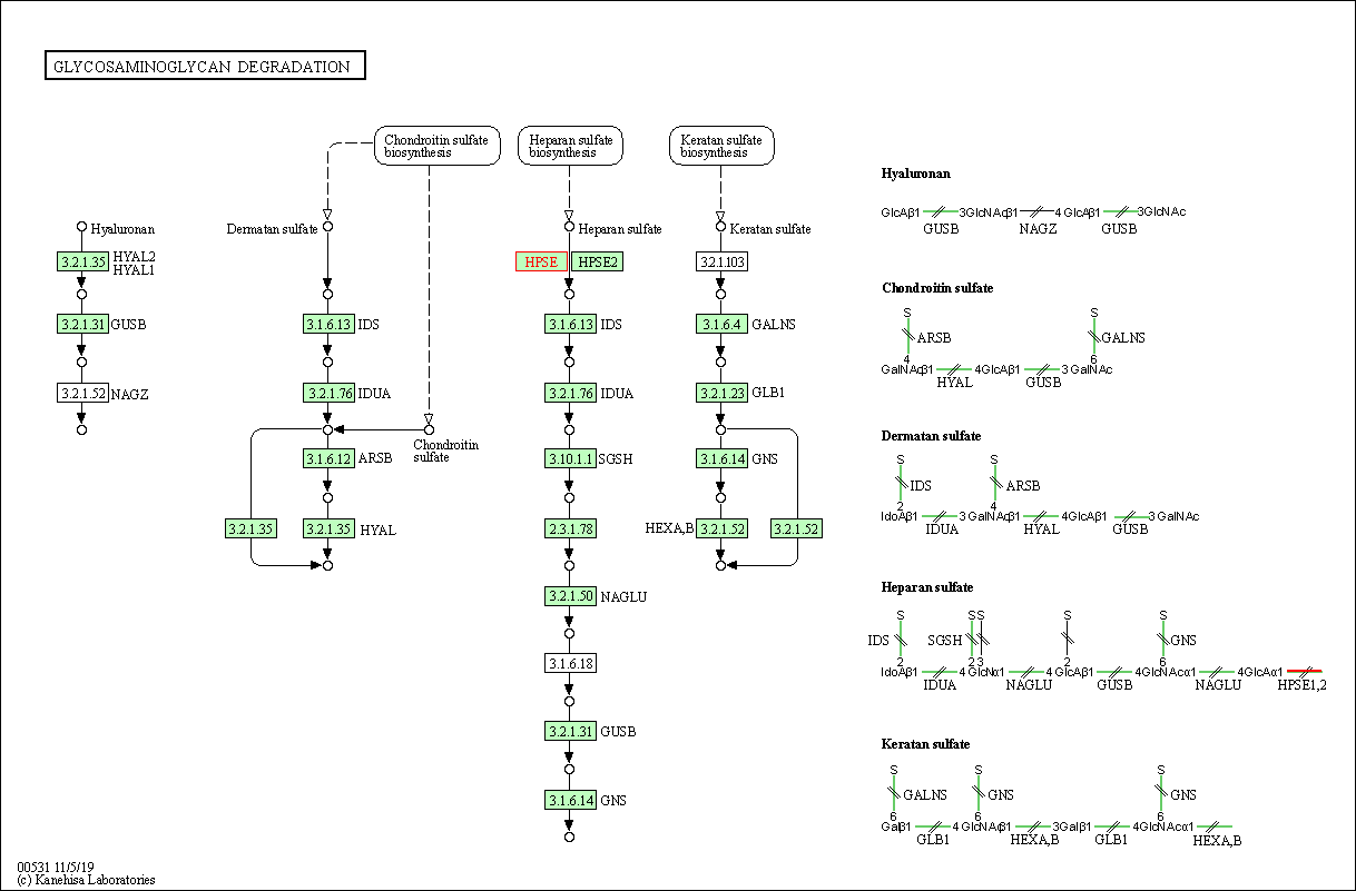
| Glycosaminoglycan degradation | hsa00531 | Affiliated Target |

|
| Class: Metabolism => Glycan biosynthesis and metabolism | Pathway Hierarchy | ||
| Degree | 2 | Degree centrality | 2.15E-04 | Betweenness centrality | 1.04E-05 |
|---|---|---|---|---|---|
| Closeness centrality | 1.99E-01 | Radiality | 1.34E+01 | Clustering coefficient | 0.00E+00 |
| Neighborhood connectivity | 2.65E+01 | Topological coefficient | 5.00E-01 | Eccentricity | 13 |
| Download | Click to Download the Full PPI Network of This Target | ||||
| Chemical Structure based Activity Landscape of Target | Top |
|---|---|
| Co-Targets | Top | |||||
|---|---|---|---|---|---|---|
| Co-Targets | ||||||
| Target Poor or Non Binders | Top | |||||
|---|---|---|---|---|---|---|
| Target Poor or Non Binders | ||||||
| Target Regulators | Top | |||||
|---|---|---|---|---|---|---|
| Target-regulating microRNAs | ||||||
| Target Profiles in Patients | Top | |||||
|---|---|---|---|---|---|---|
| Target Expression Profile (TEP) |
||||||
| Target Affiliated Biological Pathways | Top | |||||
|---|---|---|---|---|---|---|
| KEGG Pathway | [+] 3 KEGG Pathways | + | ||||
| 1 | Glycosaminoglycan degradation | |||||
| 2 | Metabolic pathways | |||||
| 3 | Proteoglycans in cancer | |||||
| PID Pathway | [+] 1 PID Pathways | + | ||||
| 1 | Syndecan-1-mediated signaling events | |||||
| Reactome | [+] 1 Reactome Pathways | + | ||||
| 1 | HS-GAG degradation | |||||
| WikiPathways | [+] 1 WikiPathways | + | ||||
| 1 | Glycosaminoglycan metabolism | |||||
| Target-Related Models and Studies | Top | |||||
|---|---|---|---|---|---|---|
| Target Validation | ||||||
| References | Top | |||||
|---|---|---|---|---|---|---|
| REF 1 | URL: http://www.guidetopharmacology.org Nucleic Acids Res. 2015 Oct 12. pii: gkv1037. The IUPHAR/BPS Guide to PHARMACOLOGY in 2016: towards curated quantitative interactions between 1300 protein targets and 6000 ligands. (Ligand id: 1728). | |||||
| REF 2 | PG545, a dual heparanase and angiogenesis inhibitor, induces potent anti-tumour and anti-metastatic efficacy in preclinical models. Br J Cancer. 2011 Feb 15;104(4):635-42. | |||||
| REF 3 | Opportunities and challenges in antiparasitic drug discovery. Nat Rev Drug Discov. 2005 Sep;4(9):727-40. | |||||
| REF 4 | Trusted, scientifically sound profiles of drug programs, clinical trials, safety reports, and company deals, written by scientists. Springer. 2015. Adis Insight (drug id 800007559) | |||||
| REF 5 | Heparanase neutralizes the anticoagulation properties of heparin and low-molecular-weight heparin. J Thromb Haemost. 2006 Mar;4(3):560-5. | |||||
| REF 6 | Structural characterization of human heparanase reveals insights into substrate recognition. Nat Struct Mol Biol. 2015 Dec;22(12):1016-22. | |||||
| REF 7 | Activity-based probes for functional interrogation of retaining beta-glucuronidases. Nat Chem Biol. 2017 Aug;13(8):867-873. | |||||
If You Find Any Error in Data or Bug in Web Service, Please Kindly Report It to Dr. Zhou and Dr. Zhang.

